Postural Control and Short-Term Adaptation Across Age and Cognitive Groups: Associations With Paratonia

Objective: To compare static balance and perturbation adaptation across young controls, cognitively healthy older adults, mild cognitive impairment (MCI), and Alzheimer’s disease (AD), and to explore whether paratonia is associated with these postural outcomes.
Methods: Fifty-four participants (19 young controls, 12 older controls, 12 MCI, and 11 AD) underwent static testing with eyes open and closed and ten backward platform perturbations. Sway path was analysed using mixed-effects models with linear and quadratic trial terms. Paratonia was assessed clinically and by surface electromyography (EMG) during passive elbow movements.
Results: Static sway differed among groups (p<.001) and increased by 8.8% with eye closure (p=.003), without an interaction. Dynamic sway also differed among groups (p<.001); values were lower in young controls than in all older groups and in MCI than in AD. Adaptation was nonlinear (p<.001) and differed among groups (p=.002). Only young controls showed a significant reduction in sway from trial 1 to trial 10 (−27.0%; p=.002), and the trajectory differed significantly only between young controls and AD (p=.004). Both EMG paratonia indices differed among groups and correlated with their corresponding clinical scores (ρ=.687 and .520; both p<.001), but neither was associated with static sway or adaptation.
Conclusion: Static and dynamic postural control varied with age and cognitive status, whereas a significant reduction in sway across repeated perturbations was observed only in young controls. Paratonia was not associated with static instability or the adaptation trajectory.
Alzheimer’s disease, balance, electromyography, mild cognitive impairment, paratonia, postural adaptation, postural control

Journal of Movement & Neuromuscular Science
Noventra Scientific Publishing
Postural Control and Short-Term Adaptation Across Age and Cognitive Groups: Associations With Paratonia
Corresponding author: Lucio Marinelli · lucio.marinelli@unige.it
Abstract
Background: Postural instability accompanies ageing and cognitive decline. Paratonia may represent a complementary motor phenotype potentially reflecting altered frontal motor control, but its relationship with postural control remains uncertain.Objective: To compare static balance and perturbation adaptation across young controls, cognitively healthy older adults, mild cognitive impairment (MCI), and Alzheimer’s disease (AD), and to explore whether paratonia is associated with these postural outcomes.
Methods: Fifty-four participants (19 young controls, 12 older controls, 12 MCI, and 11 AD) underwent static testing with eyes open and closed and ten backward platform perturbations. Sway path was analysed using mixed-effects models with linear and quadratic trial terms. Paratonia was assessed clinically and by surface electromyography (EMG) during passive elbow movements.
Results: Static sway differed among groups (p<.001) and increased by 8.8% with eye closure (p=.003), without an interaction. Dynamic sway also differed among groups (p<.001); values were lower in young controls than in all older groups and in MCI than in AD. Adaptation was nonlinear (p<.001) and differed among groups (p=.002). Only young controls showed a significant reduction in sway from trial 1 to trial 10 (−27.0%; p=.002), and the trajectory differed significantly only between young controls and AD (p=.004). Both EMG paratonia indices differed among groups and correlated with their corresponding clinical scores (ρ=.687 and .520; both p<.001), but neither was associated with static sway or adaptation.
Conclusion: Static and dynamic postural control varied with age and cognitive status, whereas a significant reduction in sway across repeated perturbations was observed only in young controls. Paratonia was not associated with static instability or the adaptation trajectory.
Keywords: Alzheimer’s disease; balance; electromyography; mild cognitive impairment; paratonia; postural adaptation; postural control
Introduction
Postural control relies on the continuous integration of sensory information and motor responses to maintain stability under changing environmental conditions [1, 2]. Ageing is commonly associated with greater postural instability and less efficient responses to unexpected perturbations [1–4]. However, poorer baseline performance does not necessarily imply a reduced ability to adapt. Repeated exposure to the same perturbation can lead to progressive modification of postural responses, and evidence suggests that at least part of this adaptive capacity may be retained in healthy older adults [5–9].
This issue becomes particularly relevant in the presence of cognitive impairment. Alterations in gait and balance have been reported in both mild cognitive impairment (MCI) and Alzheimer’s disease (AD), although their magnitude varies according to the task, sensory conditions, and outcomes considered [10–15]. Postural control also depends on cognitive resources involved in the regulation of motor responses [11, 16, 17], suggesting that cognitive decline may influence not only the magnitude of postural instability but also the way in which responses are modified across repeated perturbations. Whether short-term postural adaptation is altered in MCI and AD remains insufficiently understood.
Paratonia provides an additional perspective on motor control across ageing and cognitive decline. It is characterised by involuntary muscle activity during passive movement and may be expressed as either an oppositional response, resisting the imposed movement, or a facilitatory response, assisting it [18, 19]. Paratonia may also be observed in healthy older adults and individuals with MCI but becomes more evident clinically in dementia [18–20]. In younger individuals, related muscle activity can be detected by electromyography (EMG) even when an overt clinical manifestation is absent [19, 20]. Previous clinical and electrophysiological studies have related paratonia to altered frontal motor regulation [18–22].
Frontal cortical regions are also involved in aspects of postural control. Prefrontal activity has been observed during standing balance in young and older adults [23], while the supplementary motor area contributes to the timing of anticipatory postural adjustments [24]. These findings provide a broader neurophysiological context for examining paratonia alongside postural control, although the relationship between paratonia and static or dynamic postural performance remains uncertain.
The present study therefore compared static postural control, dynamic responses, and short-term adaptation to repeated platform perturbations among young controls, cognitively healthy older adults, individuals with MCI, and patients with AD. Clinical and EMG measures of oppositional and facilitatory paratonia were also compared across groups, and their relationships with static sway and perturbation adaptation were explored. We expected greater postural instability in the older groups and potentially smaller reductions in sway across repeated perturbations in participants with cognitive impairment.
Materials and Methods
Study design and participants
This cross-sectional observational study combined an assessment of initial static postural control with a repeated-perturbation task designed to examine short-term postural adaptation. Clinical and electromyographic measures of paratonia were also obtained to investigate their relationships with static balance and adaptation.
Participants were recruited at the Geriatric Memory Clinic, IRCCS Ospedale Policlinico San Martino, Genoa, Italy. The final stabilometric sample comprised 54 participants: 19 young healthy controls, 12 cognitively healthy older controls, 12 participants with mild cognitive impairment (MCI), and 11 patients with Alzheimer’s disease (AD).
Participants with MCI or AD underwent clinical and neuropsychological assessment as part of their diagnostic evaluation. Structural brain imaging by computed tomography or magnetic resonance imaging was considered when clinically available. Diagnoses were established according to recognised diagnostic frameworks [25, 26]. The MCI group included both amnestic and multidomain presentations. Cognitively healthy older participants had no clinical evidence of cognitive impairment, whereas young healthy adults served as an age-reference group.
Participants were required to be able to stand and walk independently and to understand and follow the experimental procedures. Exclusion criteria were parkinsonian features, a history of stroke or another acute brain lesion, polyneuropathy, and any musculoskeletal, neurological, or medical condition that prevented independent walking or the safe completion of stabilometric testing.
Clinical characterisation of the older groups included the Mini-Mental State Examination (MMSE) [27], Barthel Index (BI) [28], Instrumental Activities of Daily Living (IADL) scale [29], handgrip strength measured using a handgrip dynamometer [30], Tinetti Performance-Oriented Mobility Assessment [31], Mini Nutritional Assessment (MNA) [32], and Cumulative Illness Rating Scale–Comorbidity Index (CIRS-CI) [33]. These clinical measures were not administered to the young control group.
All participants provided written informed consent before participation. The study was conducted in accordance with the Declaration of Helsinki and was approved by the Comitato Etico Territoriale - Liguria (approval No. 15/2025; ID 14261).
Static and dynamic stabilometry
Postural control was assessed using a VertigoMed GEAHD computerised balance platform equipped with anterior and lateral safety bars. Participants stood barefoot, with their feet parallel and approximately shoulder-width apart and their arms relaxed alongside the body. The safety bars were available in the event of balance loss, and an examiner remained behind the participant throughout testing to prevent a backward fall.
Before acquisition, age, body mass, height, shoe size, and the distance between the projection of the intermalleolar line and the centre of the platform were recorded. Data were acquired at 1000 Hz per channel. The system recorded centre-of-pressure-related variables, including anteroposterior and mediolateral displacement, sway path, sway area, and sway velocity.
Total sway path, expressed in millimetres, was selected as the primary stabilometric outcome because it provides a global measure of centre-of-pressure displacement [34–36]. It was defined as the cumulative distance travelled by the centre of pressure during each acquisition epoch:
\[ \text{Sway~path} = \sum_{i = 2}^{n}\sqrt{\left( AP_{i} - AP_{i - 1} \right)^{2} + \left( ML_{i} - ML_{i - 1} \right)^{2}}, \]
where AP and ML represent the anteroposterior and mediolateral centre-of-pressure coordinates, respectively.
The stabilometric protocol comprised an initial static assessment, a dynamic perturbation task, and a final static assessment. Each static assessment consisted of ten recordings with eyes open followed by ten recordings with eyes closed while the platform remained stationary. Recordings within each visual condition were averaged to obtain one sway-path value per participant and condition. Because the present study focused on initial static postural control, only the static recordings obtained before the perturbation sequence were included in the analyses reported here.
The dynamic task comprised ten consecutive backward platform perturbations. Each perturbation consisted of a 5° backward tilt produced by inflating air cushions located beneath the right and left anterior corners of the platform to a pressure of 0.5 bar. The tilt began 0.5 s after the start of each 5-s acquisition and lasted approximately 1 s. Sway path was calculated separately over each acquisition epoch. Repeated exposure to a perturbation of identical direction and magnitude was used to examine short-term within-session modification of the postural response [5, 8].
Dynamic trial order was reconstructed from the acquisition chronology and contemporaneous operator annotations. Setup acquisitions, technical repetitions, and recordings that did not correspond to an actual perturbation were excluded, irrespective of the magnitude of the sway response. All 54 participants contributed ten perturbation trials to the primary analysis.
Trials with possible hand support, examiner assistance, stepping, or a substantial change in stance were retained in the primary analysis because these events could not always be established with certainty from the available records. These trials were flagged and excluded in a sensitivity analysis.
Postural adaptation was defined as the trial-by-trial change in sway path across repeated perturbations. A reduction in sway path was interpreted as improved short-term control of the repeated perturbation and not as evidence of retained motor learning.
Clinical assessment of paratonia
Clinical paratonia was assessed during passive elbow mobilisation, with participants seated and instructed to remain relaxed. The examiner passively moved each elbow throughout its range of motion. Facilitatory and oppositional responses were rated separately using the Paratonia Scale, ranging from 0, indicating no paratonic response, to 4, indicating an extreme response [18–20].
Assessments were performed separately on the right and left sides. Bilateral scores were averaged to obtain participant-level clinical oppositional and facilitatory paratonia scores. These scores were used to examine the correspondence between the clinical and electrophysiological manifestations of paratonia.
Electromyographic assessment of paratonia
Surface EMG was recorded from the biceps brachii and triceps brachii during passive elbow flexion–extension movements. Preamplified surface electrodes with a fixed interelectrode distance (TSD150B, Biopac Systems Inc., USA) were positioned over the muscle bellies in accordance with established surface-EMG recommendations [37–39]. Elbow excursion was monitored continuously using an electronic goniometer (TSD130B, Biopac Systems Inc., USA). Resting EMG activity was recorded for 30 s before passive mobilisation.
Passive movements were paced using a metronome and performed according to two movement patterns: continuous sinusoidal movements and discontinuous linear movements. Each pattern was performed at 60 and 100 beats/min, resulting in four experimental blocks: sinusoidal movements at 60 and 100 beats/min and linear movements at 60 and 100 beats/min. Each block comprised 15 consecutive flexion–extension cycles, following the previously described EMG paratonia protocol [19–21].
The EMG signals were visually inspected for artefacts, band-pass filtered at 20–250 Hz, and rectified. Flexion and extension phases were identified using the simultaneously recorded goniometric signal, and mean rectified EMG amplitude was calculated separately for each phase. During elbow flexion, biceps activity was classified as facilitatory and triceps activity as oppositional. During extension, triceps activity was classified as facilitatory and biceps activity as oppositional [19, 20].
For amplitude normalisation, three 5-s maximal isometric voluntary contractions were recorded for each muscle, with 30-s rest periods between contractions. Each contraction was divided into 500-ms epochs, and the highest mean EMG amplitude recorded in any epoch was used as the maximal voluntary contraction reference. Normalisation to an individual maximal reference was used to reduce between-participant variability in EMG amplitude [40].
Normalised EMG activity was calculated as:
\[ EMG_{norm} = 100 \times \frac{EMG_{mean} - EMG_{background}}{MVC}, \]
where EMGmean is the mean rectified activity recorded during the relevant movement phase, EMGbackground is the mean rectified activity recorded during the 30-s resting period, and MVC is the maximal 500-ms mean amplitude obtained during the maximal voluntary contractions.
Participant-level normalised facilitatory (EMGfacN) and oppositional (EMGoppN) indices were calculated by averaging the corresponding values across muscles, movement patterns, and movement velocities. Usable matched clinical and EMG data were available for 44 participants: 9 young controls, 12 older controls, 12 participants with MCI, and 11 patients with AD. Analyses involving EMG were conducted using these available cases.
Statistical analysis
Continuous variables were summarised as mean and standard deviation or median and interquartile range, as appropriate. Categorical variables were summarised as counts and percentages. Distributions were examined graphically and using the Shapiro–Wilk test [41].
Demographic comparisons included all four groups, whereas comparisons of geriatric and clinical measures were restricted to older controls, MCI, and AD. Overall group differences in continuous or ordinal variables were assessed using Kruskal–Wallis tests [42], and categorical variables were compared using Fisher’s exact test. Significant overall effects were followed by pairwise Mann–Whitney U tests with Holm adjustment [43].
Static sway path was log-transformed and analysed using a linear mixed-effects model that included group, visual condition, and their interaction as fixed effects, with a participant-specific random intercept [44]. Omnibus fixed effects were evaluated using Wald tests. Between-group contrasts were estimated separately for the eyes-open and eyes-closed conditions. An additional model restricted to older controls, MCI, and AD included age as a covariate. Model estimates were back-transformed and expressed as percentage differences with 95% confidence intervals.
Dynamic sway path was also log-transformed and analysed at the individual-trial level. Mixed-effects models included participant-specific random intercepts and random slopes for the linear trial term [44]. Trial number was centred at the midpoint of the perturbation sequence. Linear and quadratic trial terms, group, and the corresponding group-by-trial and group-by-quadratic-trial interactions were included as fixed effects.
Nested linear and quadratic models were fitted using maximum likelihood and compared using likelihood-ratio testing and the Akaike and Bayesian information criteria [45, 46]. The quadratic model provided the better representation of the observed trajectory and was retained for inference. Final parameter estimates were obtained using restricted maximum likelihood.
The group effect described differences in mean dynamic sway across the perturbation sequence. Within-group changes were estimated by contrasting trials 1 and 10. Overall differences in adaptation trajectories were assessed through joint tests of the group-by-linear-trial and group-by-quadratic-trial interaction terms. Pairwise trajectory comparisons were based on corresponding joint contrasts. The analysis was repeated after excluding trials flagged as potentially contaminated by hand support, examiner assistance, stepping, or changes in stance.
Overall group differences in EMGoppN and EMGfacN were assessed separately using Kruskal–Wallis tests [42], with epsilon-squared (ε²) as the effect-size measure. Pairwise percentage differences were estimated using log-linear regression models with HC3 heteroskedasticity-consistent standard errors [47]. Associations between each normalised EMG index and its corresponding clinical paratonia score were examined using Spearman rank correlations.
Associations between EMG activity and static sway were assessed using separate age- and group-adjusted log-linear regression models for the eyes-open and eyes-closed conditions. Associations with dynamic adaptation were examined using quadratic mixed-effects models containing interactions between each EMG index and both the linear and quadratic trial terms. These models included age and group as covariates and participant-specific random intercepts and linear trial slopes. The EMG-by-trial and EMG-by-quadratic-trial terms were evaluated jointly. Sensitivity analyses excluded potentially contaminated trials and repeated the dynamic models after restriction to the older groups.
Holm adjustment [43] was applied separately within each prespecified family of comparisons: post hoc comparisons for each demographic or clinical variable; six between-group contrasts within each static visual condition; six contrasts of mean dynamic sway; four within-group trial 1–10 contrasts; six between-group trajectory contrasts; six pairwise comparisons for each EMG index; four EMG–static-sway association tests; and the two EMG–adaptation interaction tests.
Estimates obtained from logarithmic models were converted to percentage differences by exponentiation. Confidence intervals for derived quantities were calculated using the delta method [48]. Missing values were not imputed. Model convergence and residual distributions were examined before inferential interpretation. All tests were two-sided, and statistical significance was set at p<.05. Analyses were performed in Python using SciPy, pandas, and statsmodels [49–51].
Results
Participant characteristics
Participant characteristics are summarised in Table 1. Participants with AD were older than older controls. Within the older cohort, participants with AD had lower MMSE, BI, IADL, Tinetti, and MNA scores than older controls. MMSE, BI, IADL, and Tinetti scores were also lower in AD than in MCI.
| Characteristic | Young controls (n=19) | Older controls (n=12) | MCI (n=12) | AD (n=11) | p value |
|---|---|---|---|---|---|
| Female sex, n (%) | 12 (63.2) | 9 (75.0) | 5 (41.7) | 7 (63.6) | .431 |
| Age, years | 30.0 [28.0–31.5] | 75.5 [73.8–79.0] | 79.0 [76.8–84.0] | 83.0 [78.5–85.0]a | <.001 |
| MMSE | — | 30.0 [29.8–30.0] | 28.0 [27.0–29.2] | 19.0 [17.0–22.0]b | <.001 |
| BI | — | 100.0 [100.0–100.0] | 100.0 [100.0–100.0] | 95.0 [82.5–100.0]b | <.001 |
| IADL | — | 8.0 [8.0–8.0] | 8.0 [7.5–8.0] | 4.0 [1.5–5.0]b | <.001 |
| HGS | — | 21.5 [20.2–30.3] | 23.6 [17.9–33.3] | 20.8 [15.7–22.7] | .811 |
| Tinetti score | — | 28.0 [28.0–28.0] | 28.0 [28.0–28.0] | 26.0 [14.5–27.0]b | .002 |
| MNA | — | 27.8 [26.9–29.2] | 25.8 [25.0–26.6] | 24.0 [22.0–25.0]a | .006 |
| CIRS-CI | — | 2.0 [2.0–4.0] | 3.0 [2.0–4.0] | 3.0 [2.0–5.0] | .811 |
Values are n (%) or median [IQR]. Overall p values for age and sex refer to all four groups; those for clinical variables refer to older controls, MCI, and AD. Clinical variables were not assessed in young controls. a Significantly different from older controls. b Significantly different from older controls and MCI. AD, Alzheimer’s disease; BI, Barthel Index; CIRS-CI, Cumulative Illness Rating Scale–Comorbidity Index; HGS, handgrip strength; IADL, Instrumental Activities of Daily Living; MCI, mild cognitive impairment; MMSE, Mini-Mental State Examination; MNA, Mini Nutritional Assessment.
Static postural control
Static postural control differed across groups (p<.001; Table 2). Overall, postural sway was 8.8% greater with eyes closed than with eyes open (95% CI 3.0–14.9%; p=.003), and this effect did not differ significantly among groups (p=.070).
With eyes open, young controls showed 30.1% lower postural sway than older controls (p=.010) and 41.5% lower sway than participants with AD (p<.001). Participants with MCI showed 28.8% lower sway than those with AD (p=.038); this difference remained significant after adjustment for age (29.9%; p=.046).
With eyes closed, young controls showed 28.7% lower postural sway than older controls (p=.017) and 32.9% lower sway than participants with AD (p=.005). No other between-group differences were significant.
| Group | Eyes open | Eyes closed |
|---|---|---|
| Young controls (n=19) | 60.9 [49.7–71.8] | 61.9 [57.2–75.7] |
| Older controls (n=12) | 80.5 [73.3–103.1] | 95.2 [78.1–102.7] |
| MCI (n=12) | 67.1 [56.8–80.7] | 77.8 [69.9–115.2] |
| AD (n=11) | 109.9 [97.3–117.9] | 102.1 [85.0–122.3] |
Values are median [IQR] sway path in millimetres; lower values indicate better static balance. AD, Alzheimer’s disease; MCI, mild cognitive impairment.
Dynamic postural adaptation
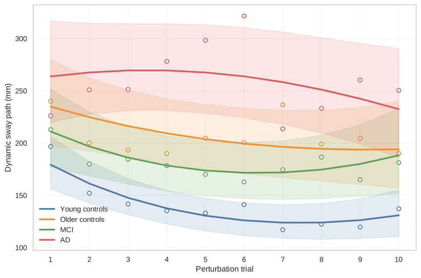
Dynamic sway differed across groups during the ten repeated perturbations (p<.001; Table 3 and Figure 1). Young controls showed 33.2% lower model-estimated dynamic sway than older controls (p<.001), 24.7% lower sway than participants with MCI (p=.010), and 46.7% lower sway than participants with AD (p<.001). Participants with MCI showed 29.2% lower sway than those with AD (p=.006). All participants were naive to the task.
The adaptation trajectory was nonlinear (p<.001), and its shape differed among groups (p=.002). From trial 1 to trial 10, sway decreased by 27.0% in young controls (p=.002), whereas the reductions in older controls (−17.4%), MCI (−10.8%), and AD (−11.9%) were not significant. The trajectory differed significantly only between young controls and participants with AD (p=.004). This difference remained significant after excluding potentially contaminated trials (p=.002).
| Group | Model-estimated dynamic sway (mm) | Trial 1 (mm) | Trial 10 (mm) | Change (%) | p for change |
|---|---|---|---|---|---|
| Young controls | 137.7 | 179.3 | 130.9 | −27.0 | .002 |
| Older controls | 206.3 | 234.9 | 194.1 | −17.4 | .291 |
| MCI | 182.9 | 211.0 | 188.3 | −10.8 | .585 |
| AD | 258.3 | 263.8 | 232.4 | −11.9 | .585 |
Values are model estimates from all 54 participants. Negative changes indicate reduced sway from trial 1 to trial 10. The p values are Holm-adjusted. AD, Alzheimer’s disease; MCI, mild cognitive impairment.
Electromyographic paratonia and postural performance
Both normalised oppositional and facilitatory EMG activity differed among groups (p<.001 and p=.006, respectively; Table 4). Oppositional EMG was lower in young than older controls (−84.5%; p=.001), MCI (−81.9%; p=.006), and AD (−91.7%; p<.001). Facilitatory EMG was similarly lower in young controls than in older controls (−87.9%; p=.006), MCI (−86.3%; p=.011), and AD (−90.6%; p=.002). No significant differences were found among older controls, MCI, and AD.
Normalised oppositional EMG was positively correlated with the corresponding clinical paratonia score (ρ=.687; p<.001). A similar correlation was found between normalised facilitatory EMG and clinical facilitatory paratonia (ρ=.520; p<.001).
Neither EMG measure was associated with static sway under eyes-open or eyes-closed conditions after adjustment for age and group (all Holm-adjusted p=1.000). Moreover, neither measure significantly modified the nonlinear adaptation trajectory across the ten perturbations (both Holm-adjusted p=.293). All interactions remained nonsignificant after excluding potentially contaminated trials and when the analysis was restricted to the older groups.
| Group or analysis | n | Oppositional EMG (EMGoppN) | Facilitatory EMG (EMGfacN) |
|---|---|---|---|
| Young controls | 9 | 0.30 [0.09–0.43] | 0.44 [0.10–1.13] |
| Older controls | 12 | 1.65 [0.95–2.45] | 3.86 [2.27–5.72] |
| MCI | 12 | 1.36 [0.53–3.39] | 2.84 [1.51–4.92] |
| AD | 11 | 2.30 [1.48–4.32] | 4.75 [2.77–5.76] |
| Overall group effect | 44 | p<.001; ε²=.362 | p=.006; ε²=.234 |
| Correlation with corresponding clinical score | 44 | ρ=.687; p<.001 | ρ=.520; p<.001 |
Values are median [IQR]. Higher values indicate greater normalised EMG activity. AD, Alzheimer’s disease; EMG, electromyography; EMGfacN, normalised facilitatory EMG; EMGoppN, normalised oppositional EMG; MCI, mild cognitive impairment.
Discussion
This study examined static postural control, dynamic responses to repeated perturbations, and their relationships with paratonia across healthy ageing, MCI, and AD. Four main findings emerged. First, static sway differed among groups and increased with eye closure, with no significant group-by-vision interaction. Second, dynamic sway was lowest in young controls and highest in AD, while participants with MCI performed better overall than those with AD. Third, adaptation followed a nonlinear trajectory: only young controls showed a significant reduction between the first and tenth perturbations, and the only significant pairwise difference in trajectory was between young controls and AD. Finally, oppositional and facilitatory paratonia differed between young participants and the three older groups and were associated with the corresponding clinical paratonia scores, but no association was detected with static balance or dynamic adaptation.
Static postural control
The lower static sway observed in young controls than in cognitively healthy older adults is consistent with the age-related decline in sensory integration and neuromuscular regulation of upright stance [1–4]. Young controls also showed less sway than participants with AD under both visual conditions. Taken together, these findings indicate that static postural control deteriorates with ageing and is particularly impaired in AD, although they do not suggest a linear progression from healthy ageing through MCI to AD.
The difference between MCI and AD reached significance only in the eyes-open condition and remained significant after adjustment for age. Greater sway in AD under this condition therefore does not appear to be explained solely by the older age of this group. However, because the group-by-vision interaction was not significant, the data do not show that the difference between MCI and AD actually varied between the eyes-open and eyes-closed conditions. The eyes-open finding should therefore be regarded as a difference identified in a specific pairwise comparison rather than as evidence of differential visual dependence between the two groups. This caution is consistent with previous research showing that the magnitude of balance impairment in MCI and AD varies according to the postural task, sensory condition, and stabilometric outcome considered [10, 12–15].
Eye closure produced a modest overall increase in sway, in keeping with the contribution of visual information to postural stability [1, 2]. This effect did not differ significantly among groups. Although the absence of an interaction should not be interpreted as evidence of equivalent responses, the present findings do not indicate disproportionate visual dependence in MCI or AD.
Dynamic postural performance and adaptation
Dynamic sway also differed substantially among groups. Young controls showed less sway than cognitively healthy older adults, participants with MCI, and patients with AD, while the MCI group showed less sway than the AD group. These findings indicate that the older groups had greater difficulty controlling whole-body displacement during platform perturbations, particularly those with AD, but do not demonstrate a uniform progression from healthy ageing through MCI to AD.
The response across trials followed a nonlinear pattern, with the largest reduction generally occurring during the initial perturbations and smaller or less consistent changes thereafter. This pattern is compatible with rapid recalibration of postural responses when the same perturbation is repeatedly encountered [5–8]. Nonlinear modelling therefore captured the full trajectory without assuming a constant rate of change across the ten trials.
Only young controls showed a statistically significant reduction between the first and tenth perturbations. Estimated reductions were also observed in cognitively healthy older adults, participants with MCI, and patients with AD, but their confidence intervals included no change. The overall trajectory differed among groups, although the only significant pairwise difference was between young controls and AD. The persistence of this finding after exclusion of potentially contaminated trials indicates that it was not driven by those trials.
Overall, these findings provide limited evidence of short-term adaptation in older adults and people with cognitive impairment. Previous studies have shown that healthy older adults can adapt to repeated slips or postural perturbations [6–9]. Differences in participant characteristics, perturbation type, outcome definition, and number of repetitions may explain why the reduction observed in cognitively healthy older adults did not reach significance in the present study. A nonsignificant within-group change does not establish a complete absence of adaptation; nevertheless, the available data do not support the conclusion that adaptation was preserved to the same extent as in young adults.
The MCI group showed an intermediate pattern in terms of dynamic sway magnitude: participants with MCI had greater sway than young controls but less than those with AD. Their adaptation trajectory, however, did not differ significantly from that of either young or cognitively healthy older adults. Within the present protocol, MCI was therefore associated with a larger postural response than that observed in young controls, without a clearly detectable difference in the adaptation trajectory. AD, by contrast, was characterised by both greater dynamic instability and a trajectory that differed significantly from that of young controls.
This pattern may reflect the task-specific nature of postural adaptation. Kannan and Bhatt reported impaired reactive stability and response scaling in MCI when perturbation intensity varied [52]. By contrast, the present protocol repeatedly applied a perturbation of the same direction and magnitude. Adapting to an increasingly predictable event may draw on different cognitive and sensorimotor resources from those required to respond to changing environmental demands. The relative preservation of some implicit and procedural motor processes in MCI and AD offers one possible framework for interpreting this distinction [53–55], although the ten-trial protocol used here cannot be equated with procedural learning.
The changes observed represent short-term adaptation within a single session. Retention, savings, transfer, and generalisation were not assessed. The findings should therefore not be interpreted as evidence of lasting motor learning or equivalent adaptive capacity across groups.
Paratonia and postural control
Oppositional and facilitatory paratonia differed among groups, with young controls showing markedly lower values than cognitively healthy older adults, participants with MCI, and patients with AD. No significant differences emerged among the three older groups. Thus, the main distinction was between young and older participants rather than a progressive increase in paratonia from healthy ageing through MCI to AD.
The positive correlations between the EMG indices and the corresponding clinical paratonia scores support the relationship between the electrophysiological and clinical manifestations of paratonia. This agreement was observed for both oppositional and facilitatory responses and is consistent with previous work describing paratonia across ageing and cognitive impairment [18–22].
Despite this correspondence, paratonia was not associated with static sway under either visual condition and did not significantly modify the trajectory of sway across repeated perturbations. These findings were consistent after excluding potentially contaminated trials and when the analyses were restricted to the older groups. Thus, within the present experimental conditions, paratonia did not account for individual differences in either static postural instability or short-term adaptation.
These findings can be considered within a broader framework of frontal motor regulation. Paratonia has been interpreted in relation to altered frontal motor regulation [18–22]. Frontal cortical regions are also involved in aspects of postural control: prefrontal cortical activity has been observed during standing balance [23], while the supplementary motor area contributes to anticipatory postural adjustments [24]. This provides a neurophysiological context in which paratonia and postural control can be considered, although the present results show that the two measures did not covary at the individual level.
The lack of association may reflect the different functional demands of the two assessments. Paratonia is elicited during passive limb movement, whereas postural control requires active whole-body regulation based on the integration of sensory information and motor responses. The two measures may therefore reflect different manifestations of motor dysfunction, even when considered within a broader framework of frontal motor regulation. Future studies combining measures of cortical activity with paratonia assessment and postural testing may clarify whether specific aspects of frontal motor regulation contribute to both phenomena.
Clinical implications
The distinction between the magnitude of dynamic sway and the adaptation trajectory may be clinically relevant. An individual may show considerable instability during perturbations without having a significantly different adaptation trajectory, as observed in participants with MCI. By contrast, AD was characterised by both greater instability and a trajectory that differed from that of young controls. Assessing a single perturbation may therefore fail to capture information provided by changes in the response over repeated trials.
Perturbation-based balance training uses repeated destabilising experiences to improve reactive control [56]. The present findings do not establish the effectiveness of this type of intervention in MCI or AD, particularly because a significant reduction in sway between the first and tenth perturbations was observed only in young controls. They nevertheless support further investigation into whether additional repetitions, graded perturbations, or longer training programmes can promote adaptation in people with cognitive impairment. Rehabilitation studies should examine retention, transfer, and effects on falls rather than assume that short-term changes observed in the laboratory translate directly into clinical benefit [57].
Interpretation is limited by the modest sample size, cross-sectional design, and age differences among groups. These age differences may have influenced some between-group comparisons, particularly those involving MCI and AD, and residual confounding cannot be completely excluded. EMG availability was also unequal across groups, with usable recordings available for 9 of 19 young controls and for all participants in the three older groups; this differential availability may limit the generalisability of EMG group comparisons. In addition, MVC-based normalisation may be influenced by age-, strength-, or cognition-related differences in the ability to generate a reliable maximal voluntary contraction. The dynamic protocol comprised only ten repetitions of the same perturbation, and total sway path did not capture directional or temporal features of postural responses. Sensitivity analyses excluding potentially contaminated trials yielded consistent results.
Conclusions
Static and dynamic postural control differed according to age and cognitive status. Young controls showed less sway than the older groups, while AD was characterised by particularly high dynamic sway and an adaptation trajectory that differed from that of young controls. Only young controls showed a significant reduction in sway between the first and tenth perturbations, whereas the estimated reductions in cognitively healthy older adults, participants with MCI, and patients with AD did not reach statistical significance.
Paratonia measured with EMG correlated with the corresponding clinical scores but was not associated with static sway or short-term adaptation to repeated perturbations.
Declarations
Author Contributions: L.M.: Conceptualization, Methodology, Investigation, Data Curation, Formal Analysis, Writing - Original Draft, Project Administration. D.D.: Methodology, Investigation, Data Curation, Formal Analysis, Writing - Original Draft. F.M.: Investigation, Writing - Review & Editing. C.T.: Conceptualization, Methodology, Writing - Review & Editing, Supervision. M.L.: Conceptualization, Methodology, Writing - Review & Editing, Supervision, Project Administration. All authors have read and approved the final version of the manuscript.
Funding: This research received no external funding.
Institutional Review Board Statement: The study was conducted in accordance with the Declaration of Helsinki and was approved by the Comitato Etico Territoriale - Liguria (approval No. 15/2025; ID 14261).
Informed Consent Statement: Written informed consent was obtained from all participants involved in the study.
Data and Code Availability: The de-identified processed datasets, complete analysis tables, and analysis code supporting the findings of this study are available from the corresponding author upon reasonable request.
Acknowledgments: The authors have no acknowledgments to report.
Competing Interests: The authors declare no commercial, financial, or other relationships that could be construed as a potential conflict of interest.
Supplementary Materials: Not applicable.
Generative AI Disclosure: Generative AI was used solely for language editing and editorial support during manuscript revision. It was not used to generate data, perform analyses, or make scientific decisions. All scientific content and the final text were reviewed and approved by the authors.